Every beat
has a direction.
A guided journey through the chambers, signals and pathways that keep blood moving.
Enter the heartNot a symbol. An organ.
Behind the familiar rhythm is a living structure with walls, vessels and a very specific shape.
A muscle with an architecture
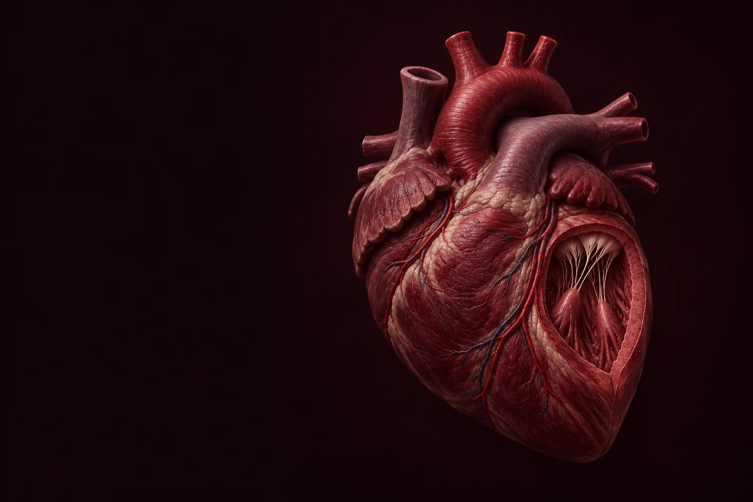
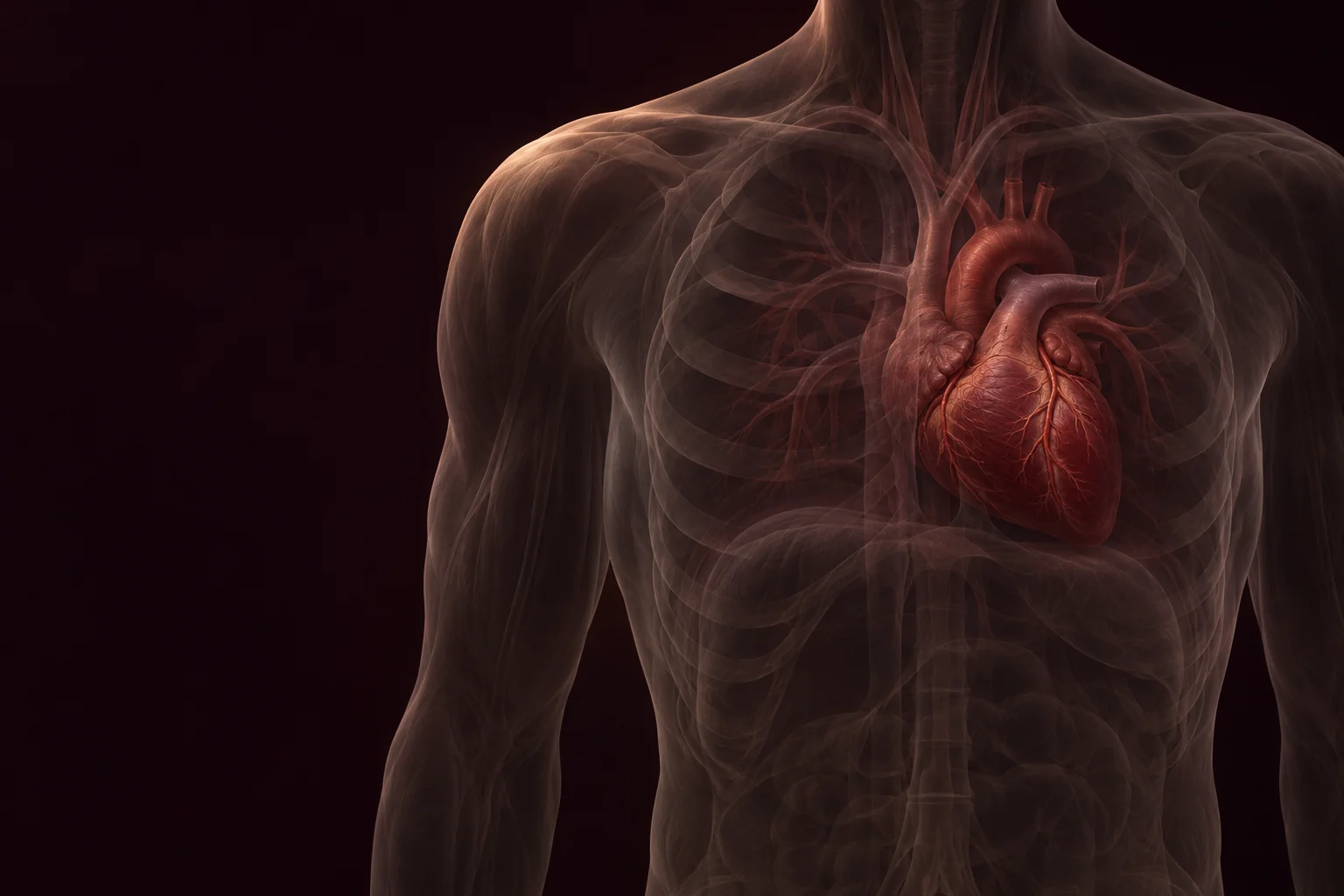
The heart sits between the lungs, with much of its mass left of the body's midline. Its irregular outline belongs to a muscular pump: receiving chambers above, pumping chambers below, and great vessels at the crown.
A surface with an inside
The myocardium forms the working muscle. A cutaway lets us look past the outer contour into the chambers while keeping the organ's orientation fixed. The shape is an illustration; the chamber relationships are the guide.
- RA — Right atrium: receives the body’s returning blood.
- RV — Right ventricle: sends blood to the lungs.
- LA — Left atrium: receives blood from the lungs.
- LV — Left ventricle: sends blood to the body.
- The septum divides the right and left sides.
Orientation
Locate the person's right and left without rotating the organ.
Locate the person's right and left without rotating the organ.
Read the diagram
- Anatomical right
- The person's right appears on your left in this anterior view.
- Anatomical left
- The person's left appears on your right in this anterior view.
Anterior orientation stays fixed throughout the heart plates. The section is an explanatory reconstruction.
Sources 01, 07Exterior to illustrated section
The Living Form
Four Chambers
Real wall edge and fixed anterior axis
Complete anatomical heart hold.
ContinueFour rooms, two directions.
The atria receive blood. The ventricles send it onward. A septum separates the sides.
- RA — Right atrium: receives the body’s returning blood.
- RV — Right ventricle: sends blood to the lungs.
- LA — Left atrium: receives blood from the lungs.
- LV — Left ventricle: sends blood to the body.
- The septum divides the right and left sides.
Name the chambers
Select a chamber to reveal its position and job.
Select a chamber to reveal its position and job.
Read the diagram
- Right atrium
- The right atrium receives oxygen-poor blood returning from the body through the venae cavae.
- Right ventricle
- The right ventricle sends blood through the pulmonary valve toward the lungs.
- Left atrium
- The left atrium receives oxygen-rich blood returning from the lungs through the pulmonary veins.
- Left ventricle
- The left ventricle sends blood through the aortic valve into the systemic circulation.
Receive above. Send below.
The right atrium receives the body's returning blood; the left atrium receives blood from the lungs. Each leads to the ventricle below it. The right ventricle serves the lungs, while the left serves the body.
A dividing wall
The atrial and ventricular septa keep the right and left sides separate in a healthy heart. The ventricular walls are muscular; the left ventricular wall is thicker than the right. The section makes these relationships visible without reversing them.
Anterior cutaway. Chamber depth and size are simplified; the septum remains visible.
Sources 01, 04, 09Electrical sequence, separate from blood flow
Four Chambers
The Signal
Right atrium and sinoatrial-node region
The chamber diagram holds.
ContinueBefore the push, a signal.
An electrical pattern coordinates atria and ventricles; it is not the path the blood takes.
- SA node — in the right atrium; starts normal activation.
- AV node — delays conduction before the ventricles.
- Dotted gold: electrical activation within heart tissue.
- Arrowed paths: blood transport, a separate process.
- 01SA originSinoatrial node
Right atrium - 02AV delayAtrioventricular node
Before ventricular activation - 03Ventricular activationConduction through
heart tissue
Body → right heart → lungs → left heart → body.
Electrical activation and blood transport are different processes, with different timescales. This is not an ECG.
Two different pathways
Compare electrical activation with blood transport.
SA node → atrial activation → AV-node delay → ventricular conduction. This is a tissue signal.
Show legend
Gold dots: electrical activation in tissue.
Solid arrows: blood through chambers and vessels.
Slate is a diagram convention. Oxygen-poor blood is dark red.
The electrical sequence and a circulation route do not share one clock.
Read the diagram
- Electrical sequence
- SA node → atrial activation → AV-node delay → ventricular conduction. This is a tissue signal.
- Blood pathway
- Blood enters chambers and leaves through vessels. It does not travel through the conduction nodes.
- Both
- The dotted signal and the arrowed blood route are separate overlays. They are not one shared clock.
Begin in the right atrium
A normal electrical impulse begins in the sinoatrial node, in the right atrium. Activation spreads through the atria and helps coordinate their contraction. These marks describe electrical activity in tissue, not a stream of blood.
A pause before the ventricles
Conduction slows at the atrioventricular node, allowing time for ventricular filling. The signal then travels through the ventricular conduction system so the ventricles can contract. This sequence and the circulation route have different meanings and timescales.
Gold dotted marks show electrical activation. Arrowed routes show circulation. The timing is explanatory, not an ECG.
Sources 03Changing explanatory layer
The Signal
The Return
Right atrial contour
The electrical overlay is visible.
ContinueA return from the body.
Blood returning from tissues enters the right atrium through large veins.
- Superior vena cava: return from the upper body.
- Inferior vena cava: return from below the heart.
- Both enter RA — the right atrium.
- Tricuspid valve: RA → RV, the right ventricle.
Trace the return
Follow the successive landmarks from the body into the right ventricle.
Systemic veins collect blood after exchange in the body's tissues.
Read the diagram
- From the body
- Systemic veins collect blood after exchange in the body's tissues.
- Venae cavae
- The superior and inferior vena cava both open into the right atrium.
- Right atrium
- The receiving chamber leads to the tricuspid valve.
- Right ventricle
- Blood crosses the tricuspid valve to enter the right ventricle.
Two major entrances
The superior vena cava returns blood from the upper body. The inferior vena cava returns blood from below. Both enter the right atrium. After supplying tissues, this returning blood contains less oxygen; it is still red.
Through the tricuspid valve
Blood passes from the right atrium through the tricuspid valve into the right ventricle. The valve sits at this chamber boundary. Its position matters: it is neither a passage to the left heart nor the outlet to the lungs.
Arrowheads indicate direction. Slate accents identify the oxygen-poor route; actual blood is dark red.
Sources 02, 04, 10Tricuspid landmark to valve comparison
The Return
One-Way Gates
Tricuspid opening between right atrium and right ventricle
The right-heart return composition holds.
ContinueDoors that answer to pressure.
Four valves open and close with changing pressure, keeping the circulation moving forward.
- Tricuspid: three leaflets; right atrium → right ventricle.
- Mitral: two leaflets; left atrium → left ventricle.
- Inlet leaflets have chordae and papillary supports.
- Pulmonary and aortic outlets: pocketlike cusps, without chordae.
Compare the valve families
Switch between the chamber entrances and ventricular exits.
Tricuspid: right atrium → right ventricle. Mitral: left atrium → left ventricle. Chordae support the leaflets.
Read the diagram
- Atrioventricular valves
- Tricuspid: right atrium → right ventricle. Mitral: left atrium → left ventricle. Chordae support the leaflets.
- Outflow valves
- Pulmonary: right ventricle → pulmonary trunk. Aortic: left ventricle → aorta. Three pocketlike cusps guard each normal outlet.
Between receiving and pumping
The tricuspid valve lies between the right atrium and ventricle; the mitral lies between the left pair. Their leaflets are supported by chordae attached to papillary muscles. These supports help keep the leaflets from turning back into the atria.
At the two exits
The pulmonary valve guards the right ventricular outlet; the aortic valve guards the left. Their pocketlike semilunar cusps close against returning blood. The inlet and outlet groups work at different phases; the four valves are not all open together.
Comparative valve plates show structure, not simultaneous opening. Outflow valves have no chordae.
Sources 02, 06, 07Right ventricle to pulmonary arteries
One-Way Gates
Toward the Lungs
Pulmonary valve root
The pulmonary valve root anchors the outflow route.
ContinueThe short journey outward.
From the right ventricle, blood crosses the pulmonary valve and travels to the lungs.
- Anatomical right lung appears on your left.
- Anatomical left lung appears on your right.
- Right ventricle → pulmonary valve → pulmonary trunk.
- Pulmonary arteries carry blood away toward both lungs.
Follow this branch
Highlight either pulmonary branch without changing flow direction.
Both pulmonary arterial branches carry oxygen-poor blood toward the lungs.
Read the diagram
- To the right lung
- Right ventricle → pulmonary valve → pulmonary trunk → right pulmonary artery.
- To the left lung
- Right ventricle → pulmonary valve → pulmonary trunk → left pulmonary artery.
- Show both
- Both pulmonary arterial branches carry oxygen-poor blood toward the lungs.
One outlet, two lungs
The right ventricle sends blood through the pulmonary valve into the pulmonary trunk. The route branches toward the right and left lungs. The vessel is an artery because it carries blood away from the heart.
Toward a smaller scale
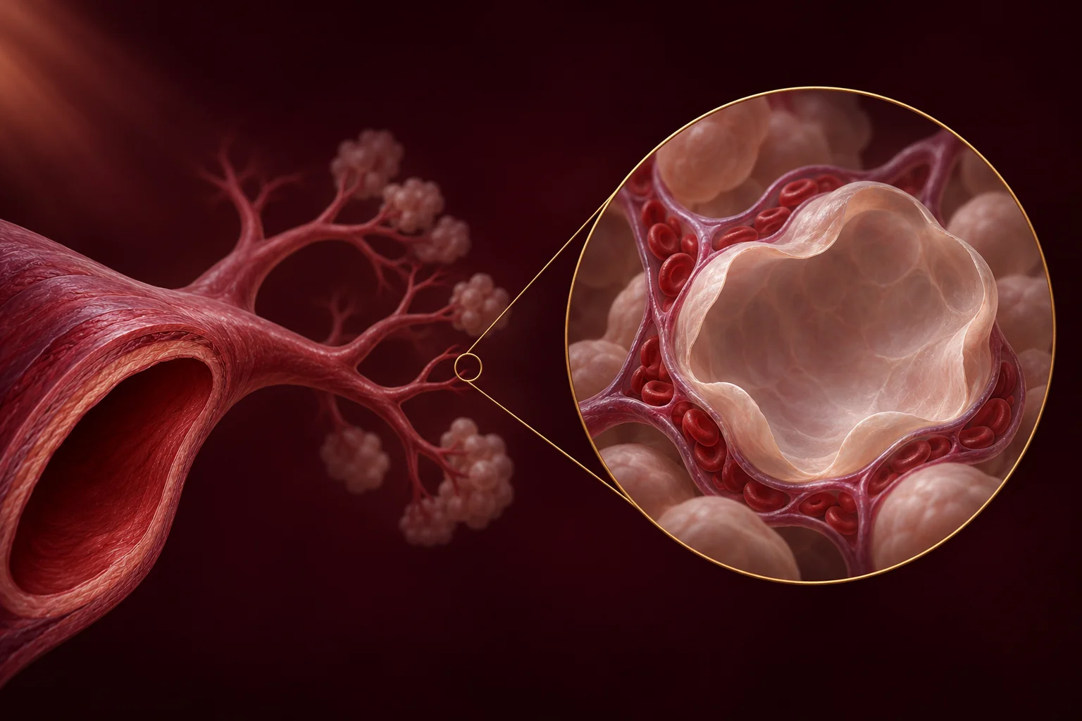
Within the lungs, the branches lead to capillaries surrounding tiny air sacs. At the next plate we change scale deliberately: a magnified diagram shows the blood vessel beside the alveolus, while keeping blood and air in separate spaces.
Schematic pulmonary route. This is oxygen-poor blood traveling away from the heart.
Sources 02, 05, 08Schematic magnification — different scale
Toward the Lungs
Exchange
Pulmonary arterial branch and explicit magnification boundary
The pulmonary artery branch is legible.
Continue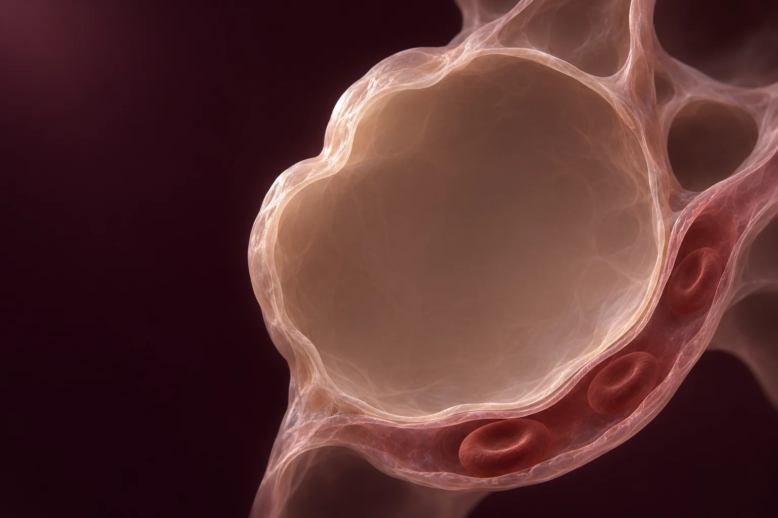
Where air meets blood—across a barrier.
Oxygen and carbon dioxide move between alveolar air and the blood through thin tissue layers.
Authored material study · not microscopy
Close, but separate
An alveolus contains air. The adjacent capillary contains blood. Their thin tissue barrier allows gases to diffuse between these compartments. Red blood cells remain inside the capillary; the alveolar space is not a blood vessel.
Two opposing directions
Oxygen moves from alveolar air into the blood, where hemoglobin carries it. Carbon dioxide moves from blood toward the air to be breathed out. The arrows show these net directions, not visible particles or a measured rate of exchange.
Air and blood remain separate. Only the gases cross the tissue barrier.
- Alveolar air is above the tissue barrier.
- Capillary blood is below; blood cells stay inside it.
- Solid O₂ arrow: oxygen moves into blood.
- Dashed CO₂ arrow: carbon dioxide moves toward air.
Gas-exchange direction
Read each arrow across the same tissue barrier.
Oxygen enters blood while carbon dioxide moves toward air. Blood and air remain physically separate.
Read the diagram
- Oxygen into blood
- Oxygen: alveolar air → tissue barrier → capillary blood.
- Carbon dioxide toward air
- Carbon dioxide: capillary blood → tissue barrier → alveolar air.
- Show both
- Oxygen enters blood while carbon dioxide moves toward air. Blood and air remain physically separate.
Schematic scale. The membrane is enlarged for legibility. This is an illustration, not microscopy.
Sources 05From microcirculation to pulmonary veins
Exchange
The Way Back
Capillary outlet and oxygen-rich notation
The alveolar diagram keeps air and blood spaces distinct.
ContinueThe return is different now.
From the lungs, pulmonary veins carry oxygen-rich blood into the left side of the heart.
- Pulmonary veins enter LA — the left atrium.
- Posterior entries are exposed in this cutaway.
- Mitral valve: LA → LV, the left ventricle.
- The separate body return enters RA — the right atrium.
Compare the two returns
Keep the same orientation while comparing the receiving chambers.
Lung capillaries → pulmonary veins → left atrium → mitral valve → left ventricle. This return is oxygen-rich.
Read the diagram
- From body
- Body tissues → systemic veins → venae cavae → right atrium. This return is oxygen-poor.
- From lungs
- Lung capillaries → pulmonary veins → left atrium → mitral valve → left ventricle. This return is oxygen-rich.
- Show both returns
- Both routes return blood to the heart. They enter different atria and carry different oxygen contents.
A vein can carry oxygen-rich blood
After exchange in the lung capillaries, blood collects into pulmonary veins and returns to the left atrium. A vein is named for its direction toward the heart, not for how much oxygen its blood contains.
The left-sided entrance
Blood moves from the left atrium through the mitral valve into the left ventricle. The two returns have different destinations: systemic veins enter the right atrium, pulmonary veins the left. The septum keeps these sides distinct.
Anterior cutaway. Pulmonary veins enter the left atrium posteriorly; their connections are exposed schematically.
Sources 02, 04, 08Mitral inflow to the left ventricle
The Way Back
The Stronger Wall
Mitral opening and left ventricular chamber rim
Pulmonary veins and the left atrium are readable.
Continue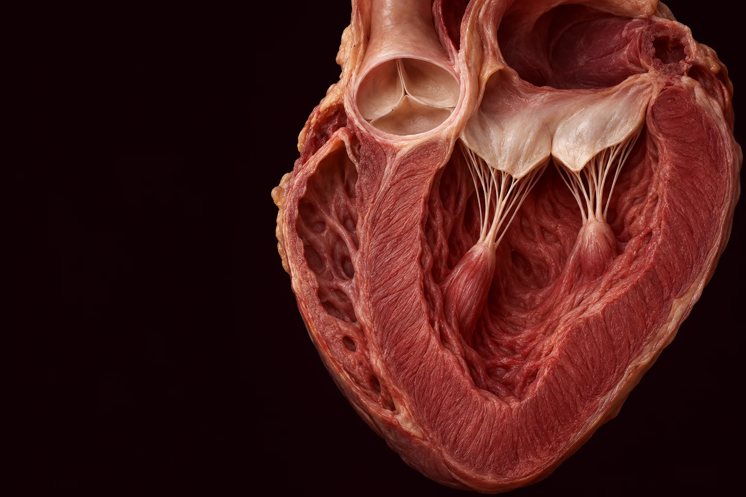
The wall built for distance.
The left ventricle has a thick muscular wall to move blood into the systemic circulation.
Authored material study · not a patient scan
Different work, different walls
The normal left ventricle has a thicker muscular wall than the right. It develops the pressure needed for the systemic circulation. The right ventricle serves the pulmonary circulation. This comparison is qualitative, not a measurement of any person's heart.
Enter through one valve. Leave through another.
The mitral valve is the inlet from the left atrium. The aortic valve is a distinct outlet into the aorta. During ventricular ejection, the mitral valve closes against backflow as blood leaves through the aortic outlet.
The muscle surrounds the chamber. Inflow and outflow remain distinct.
- RV — Right ventricular wall: thinner.
- LV — Left ventricular wall: thicker.
- Aortic outlet: left ventricle → aorta.
- This is a qualitative illustration, not a measurement.
Compare walls
Locate the two ventricular walls and then read the outlet.
The thicker left ventricular wall supports pumping into the systemic circulation.
Read the diagram
- Right ventricular wall
- The right ventricular wall is thinner in the normal heart and pumps toward the lungs.
- Left ventricular wall
- The thicker left ventricular wall supports pumping into the systemic circulation.
- Aortic outflow
- Left ventricle → aortic valve → aorta. The mitral inlet remains a separate structure.
Illustrated wall comparison. No scale bar, thickness ratio or diagnostic threshold is implied.
Sources 02, 06, 09Anatomical section to schematic body map
The Stronger Wall
Around the Body
Aortic root and aorta
The thick left ventricular wall and outlet valve hold.
ContinueOne beat, many destinations.
From the aorta, branching arteries reach tissues; blood eventually returns through veins.
- Rose arrows: aorta and arteries travel outward.
- Gold loops: tissue capillaries link arteries and veins.
- Slate arrows: veins return to the right atrium.
- This route is not a one-beat transit time.
The systemic circuit
Compare outward distribution with the return to the heart.
Arteries and veins connect through tissue capillaries. There is no direct aorta-to-vein shortcut in this normal circuit.
Read the diagram
- Arterial path
- Left ventricle → aorta → systemic arteries → tissue capillaries.
- Venous path
- Tissue capillaries → systemic veins → venae cavae → right atrium.
- Show both
- Arteries and veins connect through tissue capillaries. There is no direct aorta-to-vein shortcut in this normal circuit.
A branching distribution
The aorta receives blood from the left ventricle. Arteries and smaller branches distribute it to the body's tissues. Capillaries provide the exchange interface; the map condenses many branches into a readable route.
A return without a shortcut
Blood leaves tissue capillaries through small veins that join larger venous routes back to the right atrium. This completes the conceptual systemic circuit. Following the route here does not mean a blood cell crosses the whole body in one beat.
Schematic route, not a complete vascular atlas. Arrows indicate direction, not travel time.
Sources 02, 08Two circuits; a separate electrical sequence
Around the Body
Beat Again
Venous return entering the right atrium
The body map and returning route are readable.
Continue
The story has no final beat.
Two connected circuits and a repeating electrical pattern keep a living system moving.
Two circuits, one continuous route
The pulmonary circuit connects the right ventricle to the lungs and back to the left atrium. The systemic circuit connects the left ventricle to the body and back to the right atrium. The chambers and valves join these routes in order.
Structure carries. The signal coordinates.
Return to the same organ and choose either layer. The blood route describes where transport occurs; the electrical sequence describes how contraction is coordinated. Together they explain a heartbeat without reducing the circulation to a single moment.
The route explains where blood travels. It does not show a complete circulation in one heartbeat.
- Anatomical right appears on your left.
- Dotted gold marks show the electrical sequence.
- Arrowed paths show blood transport.
- The two layers have different meanings and timescales.
Read the atlas again
Choose the layer you want to follow, then revisit any chapter.
Dotted electrical marks and arrowed circulation paths remain separate. Neither overlay measures your heart.
Show legend
Gold dots: electrical activation in tissue.
Solid arrows: blood through chambers and vessels.
Slate is a diagram convention. Oxygen-poor blood is dark red.
The electrical sequence and a circulation route do not share one clock.
Read the diagram
- Electrical sequence
- The signal starts in the right atrium, pauses at the AV node and coordinates ventricular contraction.
- Blood pathway
- Body → right heart → lungs → left heart → body. This is a route, not a one-beat transit time.
- Both
- Dotted electrical marks and arrowed circulation paths remain separate. Neither overlay measures your heart.
The original orientation returns. Electrical and circulation overlays keep distinct meanings and timescales.
Sources 02, 03, 08